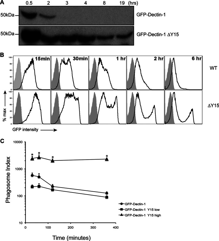
FIGURE 3.
Signaling-incompetent GFP-Dectin-1 shows enhanced retention to β-1,3-glucan-containing phagosomes. Panel A, RAW GFP-Dectin-1 (wild-type, WT) or GFP-Dectin-1ΔY15F (ΔY15) cells were incubated with β-1,3-glucan beads for the indicated time points. Following phagosome purification, recruitment of GFP-Dectin-1 WT or GFP-Dectin-1 ΔY15 was assessed by Western blot for Dectin-1 following SDS-PAGE. Panel B, GFP-Dectin-1 or GFP-Dectin-1ΔY15F recruited to purified phagosomes at various time points was determined by phagoFACS (black line, open histogram) compared with bead-only control (gray-shaded histogram). Panel C, phagosomal indices were determined from the GFP-Dectin-1 ΔY15Fhigh, GFP-Dectin-1 ΔY15Flow, and GFP-Dectin-1 WT over time. Data are representative of three independent experiments. * denotes p < 0.05.

