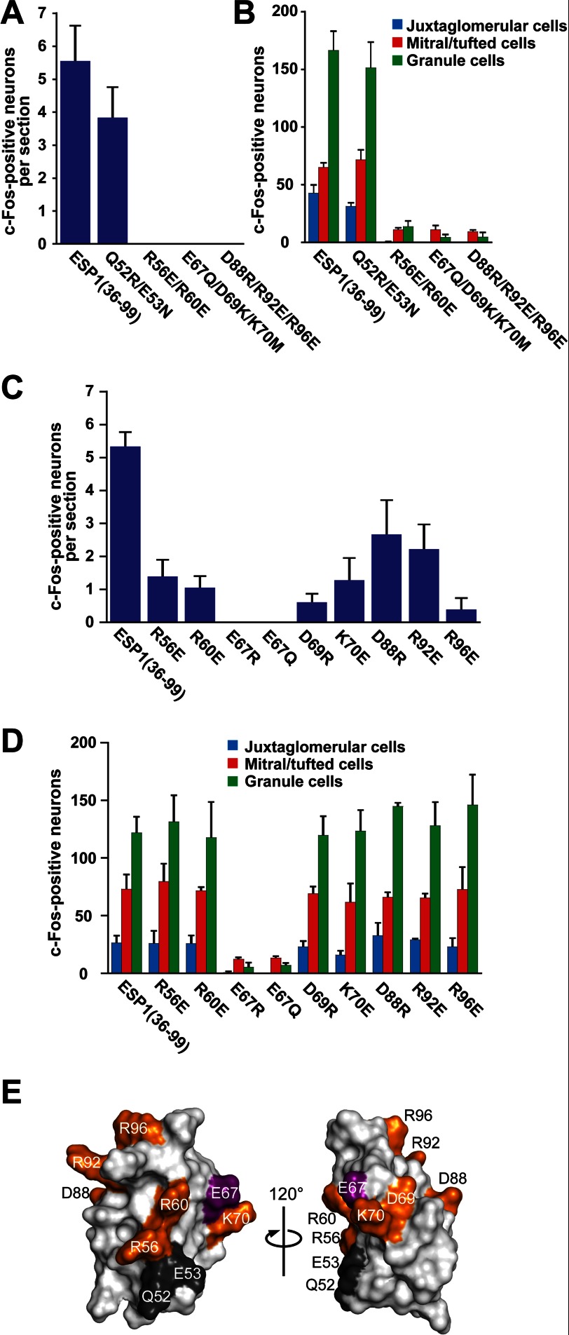
FIGURE 4.
Identification of the amino acids crucial for the c-Fos-inducing activity of ESP1. The number of c-Fos-positive VSNs was counted in BALB/c female mice stimulated with ESP1(36–99) and the multiple (A) and single mutants (C). The average values were obtained from six slice sections of the mouse VNO (n = 3 mice). The total number of c-Fos-positive AOB neurons was counted in juxtaglomerular cells (blue), mitral/tufted cells (red), and granule cells (green) stimulated with ESP1(36–99), the multiple mutants (B), or the single mutants (D) (n = 3 mice). Error bars represent S.D. E, mapping of the residues involved in the c-Fos-inducing activity of ESP1. The mutated residues for the c-Fos induction assay are colored on the ESP1 surface, where the residue crucially involved in the c-Fos inducing activity is purple, the other involved residues are orange, and the uninvolved residues are black. The two views are represented in the same manner as the left and middle panels in Fig. 3D.

