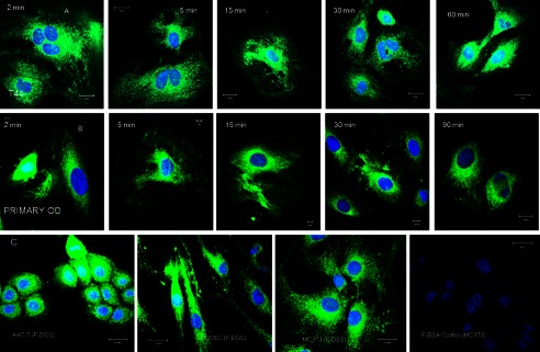
FIGURE 2.

Time course of DSS polypeptide endocytosis. A and B, representative confocal micrographs show snapshots of the location of the endocytosed FITC labeled DSS peptide (F-DSS) in T44 cells (A) and primary mouse pulp cells (B) at the indicated time points. C, representative confocal images show endocytosis of the DSS peptide by epithelial cells (HAT 7), endothelial cells (human umbilical vein endothelial cells), and osteoblast cells (MC3T3). D, shown is a negative control with FITC-labeled BSA.
