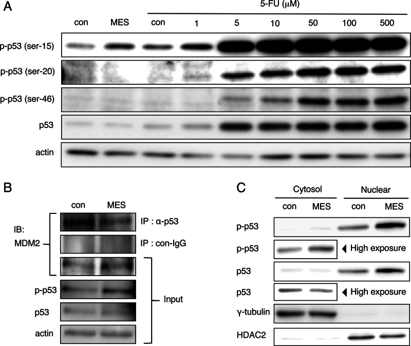
FIGURE 3.
MES enhances phosphorylation of p53 at Ser-15 and p53 recruitment to the nucleus. A, HCT116 cells were treated with MES (0.1 ms) for 30 min, and lysates were recovered 1 h after MES treatment; or cells were treated with 5-FU at the indicated concentrations for 24 h. Protein lysates were analyzed by Western blotting using the indicated antibodies. p-p53, phospho-p53; con, control. B, HCT116 cells were cross-linked 1 h after MES treatment. Protein lysates were immunoprecipitated (IP) with anti-p53 antibody (DO-1) or mouse IgG. Immunoprecipitated lysates and input samples were immunoblotted (IB) and analyzed using the indicated antibodies. C, cytosolic proteins and nuclear extract of HCT116 cells were recovered 1 h after MES treatment. Protein lysates were analyzed by Western blotting using the indicated antibodies. γ-Tubulin and HDAC2 serve as internal controls of the cytosol and nuclear fractions, respectively.

