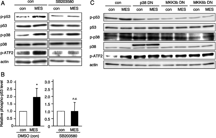
FIGURE 4.
MES induces p53 activation via the MKK3b-MKK6b-p38 pathway. A, HCT116 cells were cultured for 1 h in the presence of dimethyl sulfoxide (control (con)) or 10 μm SB203580 and then treated with MES (0.1 ms, 1 V/cm, 55 pps) for 30 min. Protein lysates were extracted 1 h after MES treatment and analyzed by Western blotting using the indicated antibodies. The experiments were performed in triplicate. p-p53, phospho-p53. B, densitometric analysis of phosphorylated p53 expression was performed using Image Gauge software. The quantified blots were normalized to actin. Error bars indicate the mean ± S.D. (n = 3). *, p < 0.05 versus the control (analyzed by Student's t test); n.s., not significant. DMSO, dimethyl sulfoxide. C, HCT116 cells were transfected with the pcDNA3.1 (control vector), p38-DN, MKK3b-DN, or MKK6b-DN plasmid. 36 h after transfection, cells were treated with MES (0.1 ms, 1 V/cm, 55 pps) for 30 min. Protein lysates were extracted 1 h after MES treatment, and Western blotting was performed.

