FIGURE 6.

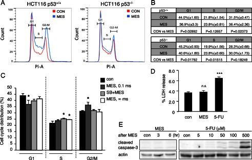
MES induces p53-dependent cell cycle arrest. A and B, HCT116 p53+/+ and HCT116 p53−/− cells were treated with MES (0.1 ms, 1 V/cm, 55 pps) for 30 min. 6 h after MES treatment, cells were washed, fixed, and stained with propidium iodide (PI). Propidium iodide-stained cells were analyzed by flow cytometry to determine cell cycle distribution. The experiments were performed in triplicate, and representative graphs are shown in A. The mean ± S.D. and p values were calculated and are shown in B. CON, control. C, HCT116 cells were pretreated with the p38 pathway inhibitor SB203580 (SB) for 1 h and then treated with MES (0.1 ms or ∞ ms) for 30 min. Cells were stained with propidium iodide 6 h after MES treatment, and the cell population was analyzed by flow cytometry. Data are presented as the percent distribution of cells in the indicated cell cycle. Error bars are the mean ± S.D. (n = 3). *, p < 0.05 (assessed by analysis of variance with the Tukey-Kramer test). D, HCT116 p53+/+ cells were treated with MES (0.1 ms) for 30 min or 5-FU (400 μm) for 24 h. LDH release was assessed 6 h after MES treatment. The experiments were performed in triplicate. Error bars indicate the mean ± S.D. ***, p < 0.001 versus the control )assessed by analysis of variance with the Tukey-Kramer test); n.s., not significant. E, HCT116 p53+/+ cells were treated with MES (0.1 ms) for 30 min, and protein lysates were isolated 3 or 6 h after MES treatment. Cells were treated with the indicated concentrations of 5-FU for 24 h, and protein lysates were recovered for analysis. Cleaved caspase-3 was detected by immunoblotting. Actin served as a loading control.
