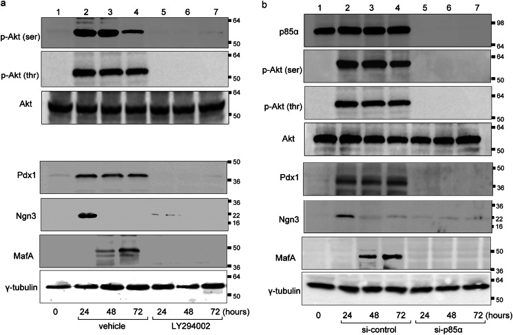
FIGURE 3.
Activation of PI3K pathway in AdAmy2TRα-infected exocrine cells. a, expression levels of phosphorylated Akt, total Akt, Pdx1, Ngn3, and MafA protein in AdAmy2TRα-infected cells exposed to 100 nm T3 for 24, 48 or 72 h with or without LY294002 treatment. b, effects of siNRA knockdown of p85α (or control siRNA) on AdAmy2TRα-infected cells incubated with 100 nm T3 for 24, 48, or 72 h and analyzed by Western blotting of cell extracts. Loading controls for γ-tubulin are shown in the bottom panel. Western blot analysis was performed with 20 μg of protein.

