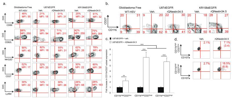
Figure 2. NK cells are activated following oHSV therapy.
(a) FACS assessment of the mean fluorescent intensities (MFI) and percentage of NK cells (CD3–DX5+) expressing various NK cell activation markers (CD69, CD62L, NKG2D, CD27, or Ly49D), 72 hours after intracranial inoculation of rQNestin34.5 into athymic mice bearing U87dEGFR human glioblastomas. Additionally, FACS quantification of the above markers is shown in KR158dEGFR syngeneic tumors and following WT HSV infection into the brains of athymic mice lacking glioblastoma (glioblastoma-free). Refer to Supplementary table 1 for average and ranges summarizing the total number of experiments. (b, c) FACS analysis of NK cell markers CD11bhighCD27high (cytotoxic) or CD11blowCD27high (immature) compared to CD11bhighCD27low (senescent), 72 hours after rQNestin34.5 or vehicle inoculation in both xenograft and syngeneic tumor models in addition to WT HSV infection into the brains of athymic mice lacking glioblastoma (glioblastoma-free). This is presented as both representative dotplots (b) and a fold increase in the expression of each NK cell population compared to vehicle treated mice (c) (n = 4–6 mice/group). (d) Percentage positivity and fold increase (in parentheses) of the degranulation marker CD107a in the CD11bhighCD27high and CD11bhighCD27low NK cell subsets 72 hours after rQNestin34.5 or vehicle treatment of mice with glioblastoma xenografts. * P < 0.05; ** P < 0.01; *** P < 0.001. Error bars represent +/− standard deviation.

