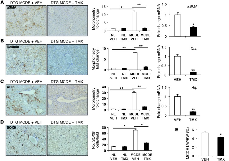
Figure 5. Blocking Hh signaling in MFs inhibits accumulation of liver epithelial progenitors, causes liver atrophy, and blocks liver cell proliferation after MCDE diet–induced injury.
(A–D) Representative immunohistochemistry, liver morphometry data, and whole liver mRNA expression for (A) αSMA, (B) desmin, (C) AFP, and (D) SOX9 in DTG mice following MCDE diet–induced injury. Mice were fed MCDE diets for 1 week, with each group receiving injections of either vehicle or TMX on days 0, 2, 4, and 6, and livers were harvested on day 7. Results are expressed either as fold over vehicle-treated sham-operated controls (computer-assisted morphometry) or total numbers of positively stained cells per field (original magnification, ×20). NL, normal. *P < 0.05; **P < 0.01. Results of qRT-PCR analysis of gene expression in the MCDE groups are shown, with results expressed as fold over vehicle-treated MCDE group. *P < 0.05; **P < 0.01. (E) Representative liver/body weight ratios, 7 days after MCDE in DTG mice treated with vehicle or TMX. Results are graphed as mean ± SEM. *P < 0.05.

