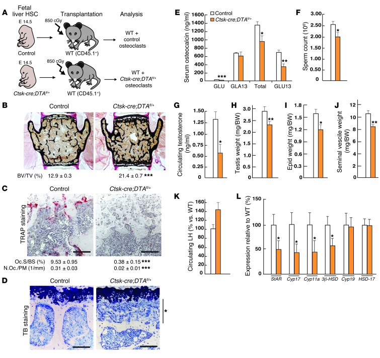
Figure 3. The osteocalcin reproductive function is hampered in the absence of proper bone resorption.
(A) Schematic representation of the strategy used to generate Ctsk-Cre;DTAfl/+ male mice. (B–D) Histological and histomorphometric analyses of vertebrae in control (n = 8) and Ctsk-Cre;DTAfl/+ (n = 9) male mice 4 months after transplantation. (B) Von Kossa/Van Giesen staining. Bone volume over trabecular volume (BV/TV%) is indicated below the pictures. (C) TRAP staining to reveal osteoclasts. Osteoclast surface per bone surface (Oc.S/BS [%]) and number of osteoclasts per bone trabecular surface (N. Oc/Bpm [1 mm]) are indicated below the pictures. (D) Toluidine blue staining showing an important presence of cartilage remnants (indicated by the asterisk) in Ctsk-Cre;DTAfl/+ male mice versus WT. (E) Measurement of uncarboxylated, carboxylated, total, and undercarboxylated forms of osteocalcin in the serum of 10-week-old Ctsk-Cre;DTAfl/+ versus WT male mice. (F) Sperm counts and (G) circulating testosterone levels; (H–J) testis, epididymal, and seminal vesicle weights normalized to BW (mg/g of BW) in Ctsk-Cre;DTAfl/+ (n = 12) versus WT (n = 11) male mice. (K) Circulating LH measurement in control and Ctsk-Cre;DTAfl/+ mice. (L) qPCR analysis of the expression of steroidogenic acute regulatory protein (StAR), cholesterol side-chain cleavage enzyme (Cyp11a), cytochrome P-450 17 α (Cyp17), 3-β-hydroxysteroid dehydrogenase (3β-HSD), aromatase enzyme (Cyp19), and 17- β-hydroxysteroid dehydrogenase (HSD-17) in Ctsk-Cre;DTAfl/+ (n = 12) compared with WT (n = 11) male mice. All analyses presented were performed on nonbreeder C57BL/6J mice. Scale bars: 200 μm). *P < 0.05; **P < 0.01; ***P < 0.001.

