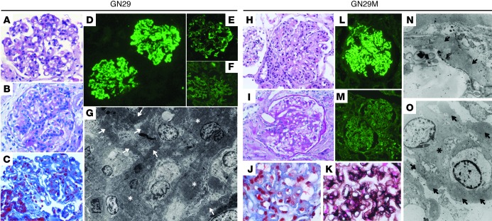
Figure 1. Histology, immunofluorescence, and EM findings.
Kidney biopsies from probate GN29 (A–G) and his mother, GN29M (H–O), showed remarkably similar light, immunofluorescence, and ultrastructural findings. The characteristic histological lesion consisted of mesangial hypercellularity with thickened, eosinophil-rich segments of GBM (A and H). The affected glomerular segments were PAS positive and reacted to trichrome and Jones methenamine-silver stain (B, C, and I–K). The main immunofluorescence findings were prominent and diffuse C3 deposits, which were granular in some glomerular areas (D and L). IgG was absent from these deposits (F and M), although local deposits of IgM were observed in GN29 (E). Both biopsies showed similar ultrastructural alterations (G, N, and O) consisting of the presence of ribbon-like, osmiophilic deposits in the GBM (arrows); these electron-dense deposits were also evident in the mesangial matrix (asterisks). Original magnification, ×400 (A–C, H, and I); ×200 (D–F); ×1,600 (G); ×600 (J and K); ×100 (L and M); ×2,950 (N); ×8,900 (O).

