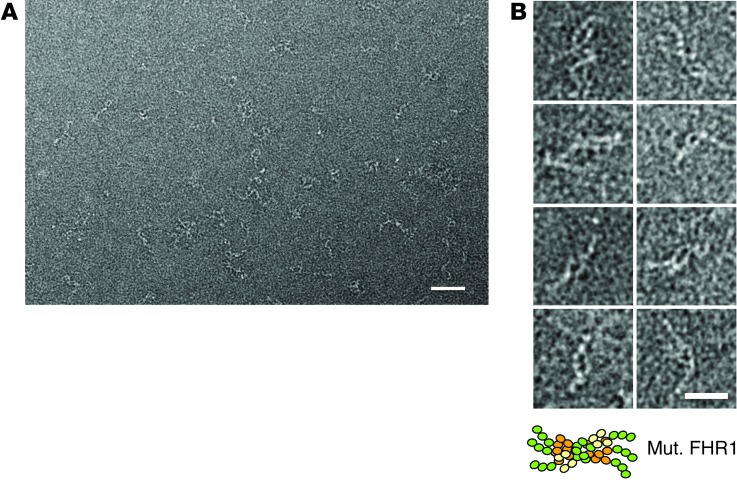
Figure 6. EM analyses of the mutant FHR1 high–molecular weight forms.
(A) Typical field for a diluted sample of a negative-staining EM analysis of the purified mutant FHR1 enriched in high–molecular weight oligomers. Isolated complexes were detected as filaments of white density on the background of the micrograph. Scale bar: 50 nm. (B) Gallery of selected images for single complexes, illustrating that each was composed of at least 2 elongated and flexible chains, presumably mutant FHR1 monomers. The length of individual chains was measured (about 30 nm) and found to be in agreement with the estimated length of an elongated mutant FHR1 molecule composed of 9 SCR domains in tandem (approximately 3 nm per SCR domain). Scale bar: 25 nm.

