Figure 1.
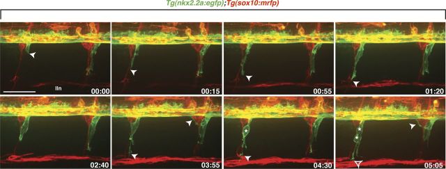
Perineurial glia migrate as a chain out of the spinal cord. Frames captured from a 24 h time-lapse movie of a wild-type Tg(nkx2.2a:egfp);Tg(sox10:mrfp) larva beginning at 48 hpf. Numbers in lower right corners denote time elapsed from the first frame of the figure. At ∼50 hpf (00:00 time point), nkx2.2a+ perineurial glia (arrowhead) exited the spinal cord next to sox10+ (red) Schwann cells. Migrating perineurial cells had very few filopodia-like projections and traveled as a chain of cells along motor nerves. Once in the periphery, perineurial glial cell bodies often divided (asterisks) and lead cells had directed membrane processes (open arrowhead) while follower cells did not. Images are lateral views of the spinal cord with dorsal to the top and anterior to the left. lin, Lateral line nerve. Scale bar, 50 μm.

