Figure 6.
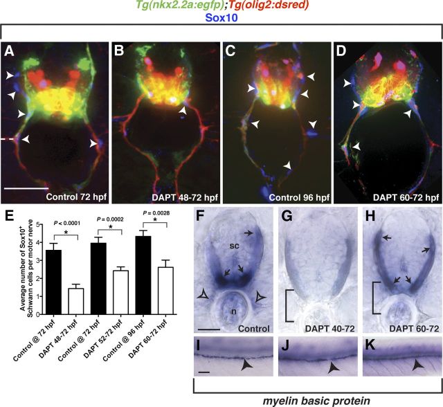
Perturbed perineurial glial development influences Schwann cell development. A–D, Transverse sections with dorsal to the top through the trunk of Tg(nkx2.2a:egfp);Tg(olig2:dsred) larvae labeled with an antibody specific to Sox10 (blue). F–K, Transverse (F–H) and whole-mount (I–K) views with dorsal to the top of mbp expression in 96 hpf larvae. A–D, Compared with controls, DAPT-treated larvae had significantly fewer Schwann cells (arrowheads) populating the motor nerve. E, Quantification of Sox10+ cells along ventral motor roots to the horizontal myoseptum. F, I, In control larvae, mbp transcript was detected in oligodendrocytes in the CNS (arrows) as well as in Schwann cells along motor nerves (open arrowheads) and the PLLn (arrowheads). G, J, In contrast, larvae treated with DAPT from 40 to 72 hpf showed strongly reduced mbp expression in the CNS and no detectable expression along motor nerves. However, PLLn expression was indistinguishable from controls. H, K, Similar to the early DAPT treatment, larvae exposed to DAPT from 60 to 72 hpf showed no motor nerve mbp expression, but indistinguishable expression in the CNS and along the PLLn. Statistical significance was determined using the unpaired t test. p-values are shown for each treatment type compared with controls. Horizontal dashed line denotes horizontal myoseptum. Scale bars: A–D, 25 μm; F–H, 25 μm; I–K, 50 μm.

