Figure 3.
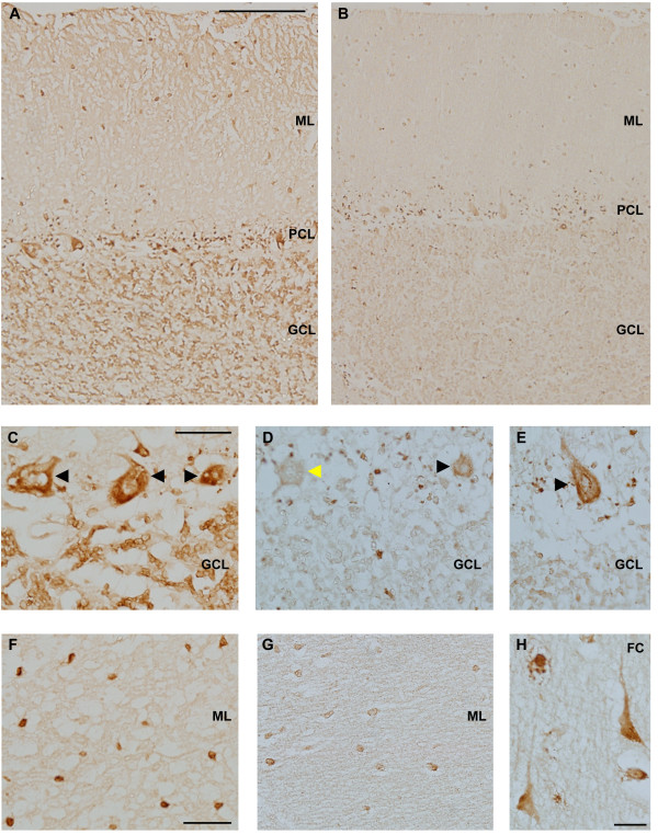
Immunohistochemistry demonstrates reduced FMRP levels in the cerebellum and frontal cortex of the proband. Paraffin embedded tissue sections from the proband and controls were immunostained with anti-FMRP (clone 1C3) antibody. (A,B): A comparison of FMRP immunoreactivity (FMRP-IR) in the cerebellar cortex of SK relative to age-matched control tissue demonstrates reduced FMRP-IR in the molecular layer (ML), purkinje cell layer (PCL) and granule cell layer (GCL). (C) FMRP immunoreactive purkinje cells present in cerebellar cortex of a control subject. The granule cell layer is characterized by significant FMRP expression in control sections which is dramatically reduced in the proband. (D, E) FMRP-positive (black arrowhead) and FMRP-negative Purkinje cells (yellow arrowhead) were present in the cerebellum of SK. (F) FMRP-positive cells in the molecular layer of cerebellar cortex of a control section. (G) Levels of FMRP are significantly reduced in the molecular layer of SK. (H) FMRP-positive cells with the morphology of excitatory neurons in the frontal cortex (FC) of SK. Scale bar in A = 200 μm, applies to A,B. Scale bar in C = 50 μm, applies to C-E. Scale bar in F = 50 μm, applies to F,G. Scale bar in H = 25 μm.

