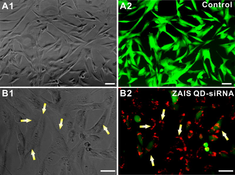
Figure 5. In vitro testing of the ZAIS-QD-siEGFP cell uptake and silencing efficiency in stably transfected U87-EGFP glioblastoma cells.

(A) Control U87-EGFP cells with PEI-coated ZAIS-QD; (A1) represents the phase contrast image and (A2) is the corresponding fluorescence image. (B): EGFP knockdown using the ZAIS QD-siRNA constructs; (B1) Phase contrast image showing the the viability of U87-EGFP cells has not changed appreciably after the transfection of the ZAIS QD-siRNA constructs as compared to the control cells in (A). (B2) Fluorescence image clearly shows the knockdown of EGFP in cells which have internalized the siRNA-QDs (red) after 72 hrs. The red fluorescence from the ZAIS QDs correlates well with the loss of the green fluorescence in cells (indicated by yellow arrows). Scale bar is 50 μm
