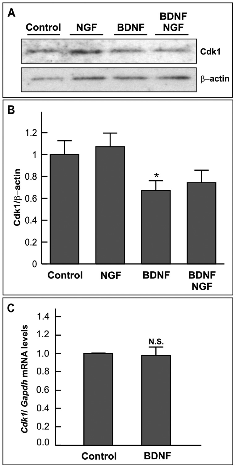
Figure 4. BDNF reduces cdk1 protein levels in DCRNs.

(A) Lysates from DCRNs cultured for 20 h in the presence of different combinations of 100 ng/m NGF and 2 ng/ml BDNF were subjected to western blot with antibodies specific for either cdk1 (upper panel) or β-actin (lower panel). (B) Normalized cdk1/β-actin ratio. (C) Semiquantitative RT-PCR analysis of mRNA samples obtained from DCRNs cultured for 20 h in the presence of either vehicle (Control) or 2 ng/ml BDNF (BDNF). The levels of Cdk1 expression were normalized to Gapdh. N.S. non-significant; *p<0.05 (Student’s t test; n = 3–4).
