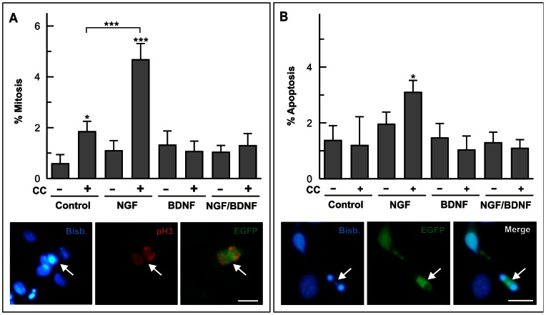
Figure 6. Post-transductional effects of BDNF on cdk1 function.
(A) E6 retinal cells electroporated with either EGFP (-) or cdk1 plus cyclin B1 (CC) and EGFP (+) were cultured under neurogenic conditions for 20 h in the presence of different combinations of 100 ng/ml NGF and 2 ng/ml BDNF. The percentage of mitotic figures was evaluated in the EGFP-positive cells. Lower panels show an example of a mitotic figure (pH3; red) in an EGFP-transfected cell (EGFP) (arrow). (B) E6 retinal cells were electroporated and cultured as above. The percentage of pyknotic nuclei was evaluated in the EGFP-positive cells. Lower panels show an example of a pyknotic nucleus in an EGFP-transfected cell (EGFP) (arrow). Bisb.: bisbenzimide. *p<0.05; ***p<0.005 (Student’s t test; n = 4). Bars: 10 µm.

