
Figure 3.
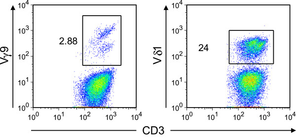
Expanded γδ T cells in the patient’s blood are Vδ1+ γδ T cells. Further flow cytometric characterization of donor γδ T cells recovered from the patient 9 months after transplantation. Ficoll-purified blood samples were stained with PE-Cy7-conjugated anti CD3 (BD bioscience, clone SK7), PC5-conjugated Vδ9 (Beckman Coulter cat. # PNA63663), and FITC-conjugated Vδ1 (Thermo Scientific, clone TS8.2, cat. # EN-TCR2730). Numbers next to quadrants indicate percentage of gated Vδ9+ (left panel) or Vδ1+ (right panel) γδ T cells among all CD3+ lymphocytes.