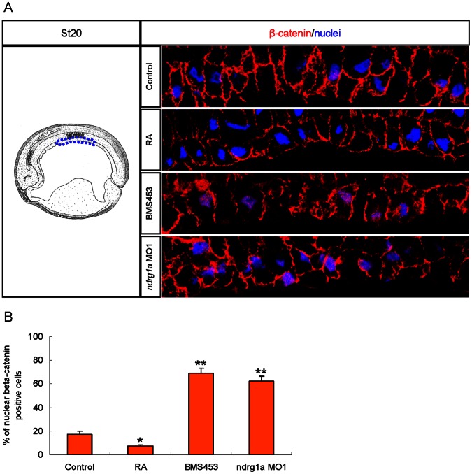
Figure 6. Nuclear β-catenin localization in archenteron roof endoderm cells appears to be suppressed by RA activated Ndrg1a in Xenopus laevis.
Embryos were treated with 5 µM RA, 0.25 µM BMS453 for one hour at stage 10, or vegetally injected with 3.5 picomoles of ndrg1a MO1 at four cell stage and collected at stage 20 for immunofluorescence. (A) Left panel is a schematic drawing illustrating a midsagittal section of stage 20 embryos (after Hausen and Riebesell [71]). The dashed blue lines outline the archenteron roof endoderm where ndrg1a is expressed. Right panels are representative immunofluorescence images showing β-catenin signals (red channel) and DAPI staining (blue channel) in the outlined archenteron roof endoderm cells. (B) Quantification data obtained from three independent experiments. Nine embryos in total (three for every experiment) from each group were sectioned to evaluate the mean percentage of β-catenin positive cells in the outlined archenteron roof endoderm illustrated in the left panel of A. For each embryo, the outlined archenteron roof endoderm cells in the 30 consecutive parasagittal sections central to the median plane were scanned for nuclear β-catenin signals. *, p<0.05. **, p<0.01 (Student’s t-test, two-tailed distribution).

