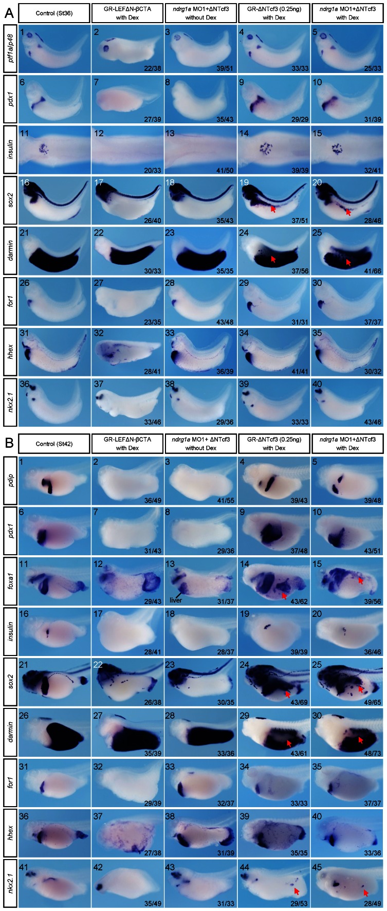
Figure 7. Ndrg1a represses Wnt/β-catenin signaling, thus specially allowing pancreas, oesophagus, stomach, and duodenum specification.
(A, B) Xenopus laevis embryos were vegetally injected with the reagents indicated on the top, treated with 10 µM dexamethasone (Dex) at stage 15, collected at stages 36 (A) and 42 (B), and subjected to whole mount staining with probes indicated on the left side. Doses of the reagents injected are as follows: ndrg1a MO1, 3.5 picomoles; GR-ΔNTcf3 mRNA, 0.25 ng; GR-LEFΔN-βCTA mRNA, 0.5 ng. Red arrows in images A19, 20, 24, 25, B14, 15, 24, 25, 29, 30, 44, and 45 point to either ectopic or loss of expression of marker genes indicated upon inhibition of Wnt/β-catenin signaling. (A11–15) Dorsal view. The dorsal structures, such as the neural tube, notochord, and somites were removed after whole mount in situ hybridization. All the rest images in A and B are lateral view with head toward the left. The numbers of embryos showing the illustrated phenotypes are given in the corresponding images.

