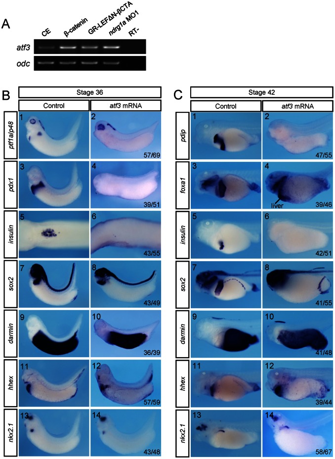
Figure 8. Overexpression of Atf3 phenocopied ndrg1a knockdown.
(A) Xenopus laevis embryos were vegetally injected with 0.25 ng of β-catenin mRNA, 0.5 ng of GR-LEFΔN-βCTA mRNA or 3.5 picomoles of ndrg1a MO1 at 4-cell stage, treated with 10 µM Dex at stage 11, and subjected to RT-PCR analysis of atf3 expression at stage 30. Ornithine decarboxylase (odc) was used as the RNA loading control. (B, C) Xenopus laevis embryos were vegetally injected with 0.3 ng of atf3 mRNA at 4-cell stage and collected at stages 36 (B) and 42 (C) for whole mount staining with probes indicated on the left side. (B5, 6) Dorsal view. The dorsal structures, such as the neural tube, notochord, and somites were removed after whole mount in situ hybridization. All the rest images in B and C are lateral view with head toward the left. The numbers of embryos showing the illustrated phenotypes are given in the corresponding images. Abbreviation: CE, control embryos.

