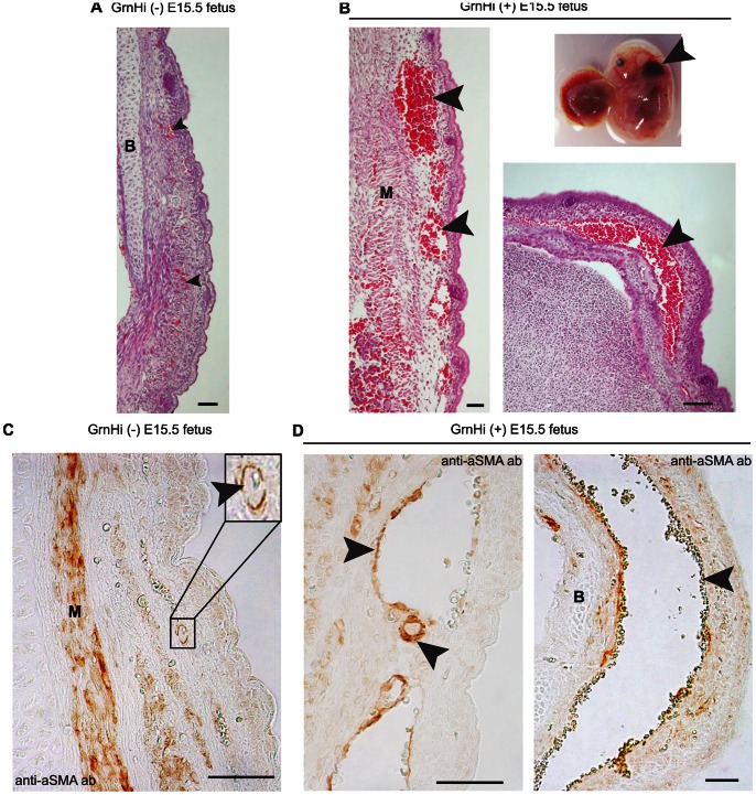
Figure 7. Compromised subcutaneous vessel structure at E15.5 in the Tie2-Grn positive embryos.
Some Tie2-Grn positive fetuses exhibited subcutaneous hemorrhage at E15.5, particularly in the head region. (A) The scalp from a Tie2-Grn negative littermate and (B) from a Tie2-Grn positive littermate showing highly enlarged vessels not present in the negative counterpart. Sections were stained with H&E, the arrow heads point to blood vessels; the insert shows the external appearance of the Tie2-Grn positive fetus with the arrow head showing the extent of subcutaneous hemorrhage. The small vessels of the Tie2-Grn negative fetus (C) are enclosed in smooth muscle α-actin positive mural cells (insert). (D) The smaller vessels in the Tie2-Grn positive fetus is enclosed smooth muscle α-actin positive mural cells but the more enlarged vessels show incomplete association with smooth muscle α-actin positive mural cells (arrow heads indicate vessels). B, bone; M, muscle. Scale bars denote 20 µm. Images were obtained from the GrnHi line.

