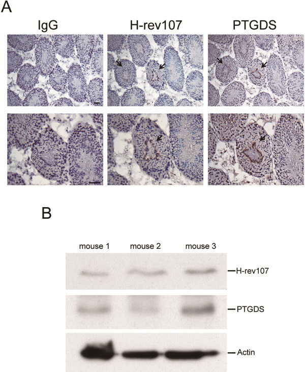
Figure 1.
Expression of H-rev107 and PTGDS in normal testis tissues. Mouse testis tissues were analyzed for expression of H-rev107 or PTGDS by immunohistochemical staining using rabbit IgG, H-rev107, or PTGDS antibody. Arrows indicate positive H-rev107 and PTGDS staining (magnification ×100, top panel; ×200, bottom panel) (A). Total cellular extracts from testis of mice were subjected to Western blot analysis for H-rev107, PTGDS, and actin (B). Scale bar: 50 μm.

