
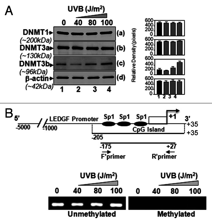
Figure 4. LECs exposed to UVB displayed enhanced expression of DNA methyl transferase DNMT3b, but could not methylate LEDGF promoter. (A) Nuclear extract from UVB exposed hLECs were resolved on 4–20% SDS gel and immunoblotted using anti-DNA methyl transferases as shown. The same membranes were used to re-probe the antibodies following restriping. Protein band appearing by β-actin antibody served as internal/loading control. Right panel shows relative densitometry of protein bands. (B) MSP analysis disclosed that status of LEDGF promoter (-175/+27) containing Sp1 sites within the CpG island was not altered. Top panel shows the diagrammatic representation of the LEDGF promoter, predicted CpG island and regions where the PCR primers bind. Lower panel shows the representative gel image from MSP analysis of genomic DNA isolated from LECs exposed to variable doses of UVB-exposed or unexposed to UVB.