
Figure 2. SPP1 expression in the motor cortex of the marmoset and squirrel monkey, and the brainstem and spinal cord of New World monkeys.
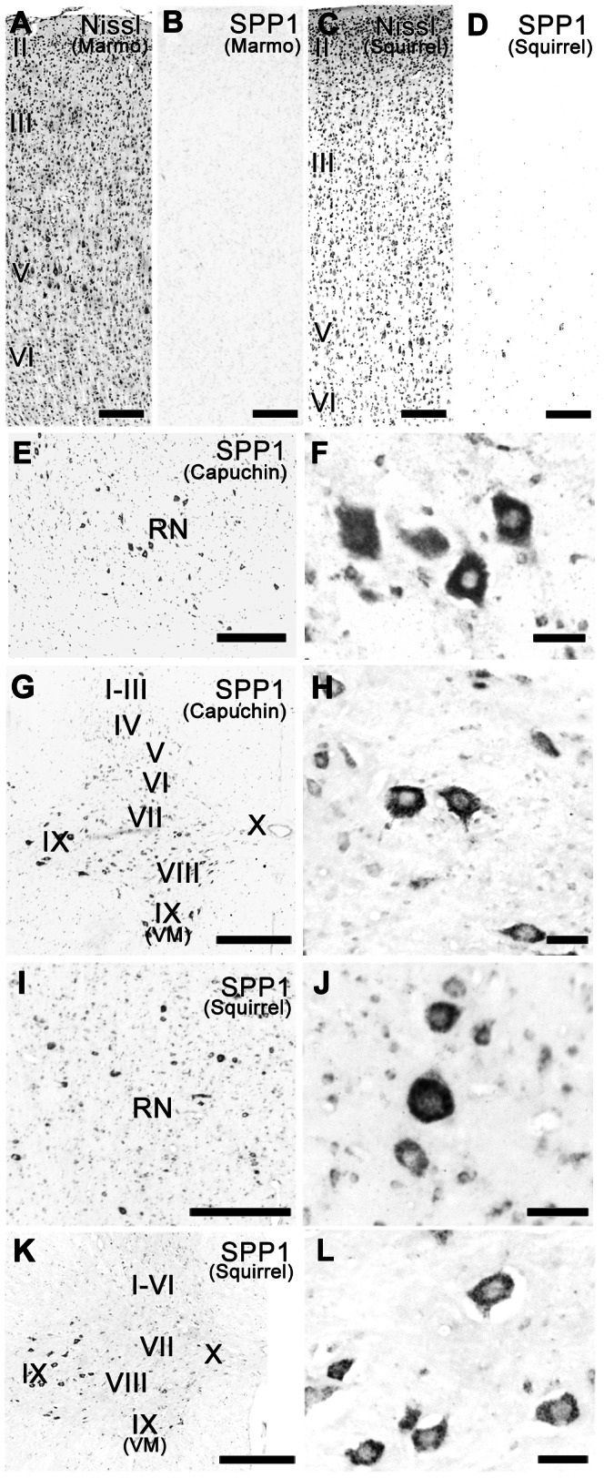
A, B: Nissl-stained (A) and adjacent sections of the marmoset primary motor cortex (M1) hybridized with the SPP1 antisense probe (B). C, D: Nissl-stained (C) and adjacent sections of the squirrel monkey M1 hybridized with the SPP1 antisense probe (D). E, F: Low- (E) and high- (F) magnification photomicrographs of SPP1 mRNA-positive neurons in the red nucleus of the capuchin monkey. G, H: Low- (G) and high- (H) magnification photomicrographs of SPP1 mRNA-positive neurons in the eighth cervical spinal segment of the capuchin monkey. I, J: Low- (I) and high- (J) magnification photomicrographs of SPP1 mRNA-positive neurons in the red nucleus of the squirrel monkey. K, L: Low- (K) and high- (L) magnification photomicrographs of SPP1 mRNA-positive neurons in the eighth cervical spinal segment of the squirrel monkey. RN, red nucleus. II–VI, layers II–VI of M1. I–X, layers I–X of the spinal cord. VM, ventral medial nucleus. Capuchin, capuchin monkey. Marmo, marmoset. Squirrel, squirrel monkey. Scale bars = 200 µm in A–D; 500 µm in E, G, I, K; 50 µm in F, H, J, L.