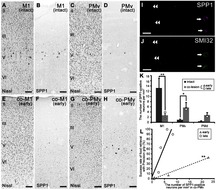
Figure 6. Changes of SPP1 expression after lesion of the lateral corticospinal tract.
A–H: Nissl-stained sections and adjacent sections showing the distribution of SPP1 mRNA-positive neurons in the primary motor cortex (M1; A, B) and ventral premotor cortex (PMv; C, D) of the intact monkey, and in the contralesional M1 (co-M1; E, F) and PMv (co-PMv; G, H) of the lesioned monkey (early stage). II–VI, layers II–VI of the cerebral cortex. I, J: Double-labeling study with SMI 32, an antibody against non-phosphorylated neurofilament H. I: Localization of SPP1 mRNA–positive neurons in layer V of the lesioned monkey (late stage). J: SMI 32 immunoreactivity in the same section as (I). Arrows indicate SPP1 mRNA–positive neurons showing SMI 32 immunoreactivity. Double arrowheads indicate SMI 32-immunoreactive neurons with no SPP1 expression. K: A bar chart showing the average density of SPP1 mRNA–positive neurons in M1, PMv, and dorsal premotor cortex (PMd) of an intact monkey, and in the co-M1, co-PMv, and contralesional PMd (co-PMd) of the lesioned monkey, with standard error. *P<0.05, **P<0.01, according to the Mann-Whitney U test. L: Scattergram showing the relationship between food retrieval success rate with precision grip and the density of SPP1 mRNA–positive neurons in the co-PMv. **P<0.01, according to linear regression analysis. The dashed and solid lines are linear approximations of the data represented by triangles and circles, respectively. The triangles and circles are data points from each lesioned monkey at early and late recovery stages, respectively. Scale bars = 200 µm in A–H; 50 µm in I, J.

