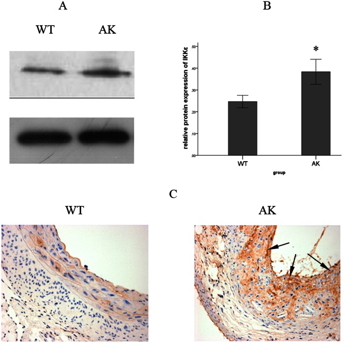
Figure 1. Protein expression and localization of IKKε after a HFD feeding.
(A) IKKε was detected by Western blotting. (B) IKKε protein expression was normalized to GAPDH in the aortic vessel wall. Expression of IKKε was notably higher in the AK group than in the WT group. Values are means ± SD; n = 9 per group. Densitometric data are from one representative experiment of three separate experiments. *P<0.05. (C) Immunohistochemistry staining of IKKε also showed that the HFD-induced expression of IKKε was mostly distributed in the intima area of the aortic vessel wall (arrows, 400×).

