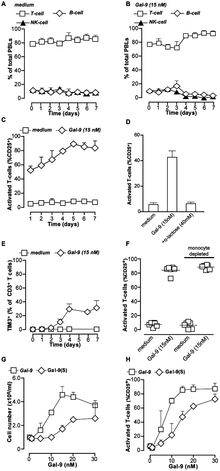
Figure 2. Recombinant and Gal-9(s) dose dependently activate T-cells.
A. Resting PBMCs (n = 5) were incubated in medium for up to 7 days. Distribution of cell populations was analyzed every day by flow cytometry. B. Similar as (A), but in the presence of 15 nM Gal-9. C. Resting PBMCs (n = 5) treated with medium or 15 nM of recombinant Gal-9 for up to 7 days were analyzed for T-cell activation by staining for activation marker CD25. D. Resting PBMCs were treated as in (C) in the presence of α-lactose, after which CD25 expression was analyzed at 1 day. E. Resting PBMCs (n = 5) were treated as in (C) and analyzed for expression of TIM-3. F. PBMCs or monocyte-depleted PBMCs were treated with Gal-9 for 7 days, and analyzed for CD25 expression G. Resting PBMCs (n = 6) were treated with a concentration range of recombinant Gal-9 or physiologically occurring isoform Gal-9(S) for 7 days and analyzed for cell density. H. Resting PBMCs (n = 6) were treated as in G and CD25 expression was determined. All graphs represent mean +/– SD.

