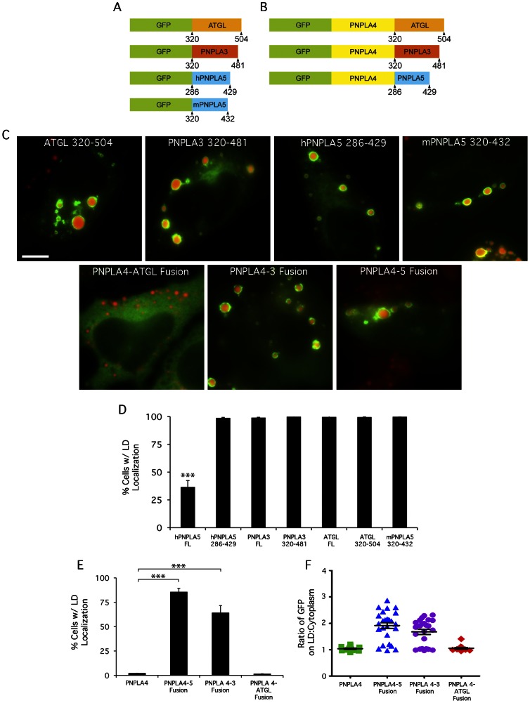
Figure 3. The C-terminal domains of other PNPLA family members are important for LD localization.
(A, B) Schematic depicting PNPLA family C-terminal domains N-terminally fused to GFP or PNPLA4. HeLa cells were treated overnight with OA, transfected with the indicated constructs for 24 h, fixed and stained with LipidTOX Red, and analyzed by fluorescence microscopy to observe LD localization. (C, top panels) PNPLA family C-terminal domains fused to GFP localize to LDs. (C, bottom panels) Fusing C-terminal domains of PNPLA3 and PNPLA5, but not ATGL, confers LD localization to PNPLA4. The number of cells with LD surface localization in a given population was quantified as a percentage of GFP-transfected cells by scoring cells that displayed ‘rings’ of GFP signal surrounding Lipidtox stained LDs. Cells lacking LDs were not scored. To account for inherent variations in LD size/number per cell, ≥300 cells were scored per condition in three independent experiments. These results, quantified in D and E, are plotted as means ± SE (≥3 experiments/condition, ≥300 cells counted/experiment, ***, p<0.0001). (F) Quantitation of fluorescence intensity on LD surface:cytoplasm ratio from line plots (n≥15 LDs and cells/condition). FL = full length, Bar, 5 μm.

