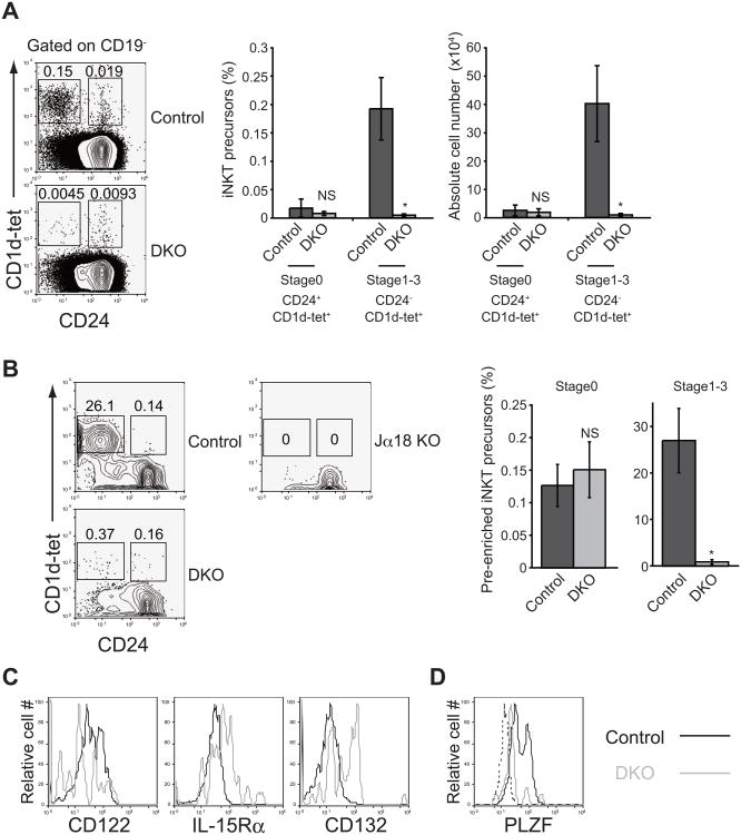
Figure 3. Loss of store-operated Ca2+ entry results in impaired iNKT cell development due to reduced expression of CD122 and PLZF.
(A) Flow cytometric analysis of α-galactosylceramide-loaded CD1d tetramer-positive iNKT cell precursors in the thymus isolated from control and DKO mice (left), frequency of iNKT cell precursors in CD19- thymocytes (center) and absolute cell number (right) of iNKT cell precursors in the thymus. Control, Stim1fl/flStim2fl/fl or Stim1+/+Stim2+/+ Vaν-iCre; DKO, Stimffl/flStim2fl/fl Vaν-iCre. n=5. (B) Flow cytometric analysis (left) and frequencies (right) of pre-enriched thymic iNKT precursors by α-galactosylceramide-loaded CD1d tetramer. Tcraj-18−/− mice (Jα18 KO) were used as a negative control. Control, Stim1fl/flStim2fl/fl or Stim1+/+Stim2+/+ Vaν-iCre; DKO, Stim1fl/flStim2fl/fl Vaν-iCre. n=5. (C) Expression of IL-15 receptor components in iNKT cell precursors from control (black lines) and DKO (grey lines) mice. (D) PLZF expression in thymic iNKT cells at stage1-3. Control: black line, DKO: grey line, Isotype control: dashed line. *, P<0.01; NS, not significant. Data are representative of two (B to D) and four (A) independent experiments. Error bars (A and B) denote mean ± SEM.

