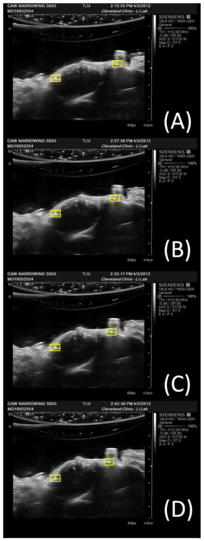
Figure 2.

Ultrasound images of a representative specimen at (a) its initial CAW of 26.7 mm, (b) 1 mm narrowing of the CAW, (c) 2 mm narrowing of the CAW, and (d) 3 mm narrowing of the CAW. The yellow boxes are the regions of interest with their centroids at the most palmar aspect of the hamate and the trapezium.
