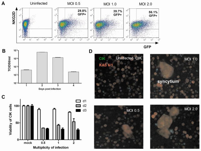
Figure 1. Infection of CIK cells with measles virus.
(A) Dual color flow cytometry showing NKG2D expression in MV-GFP infected CIK cells. Quantitation of MV infection in CIK cells was performed by flow cytometry for GFP positive cells at 24, 48 and 72 h post infection at various multiplicities of infection (MOI, ratio of virus:cell). (B) Replication of MV-Luc in CIK cells as measured by amount of progeny virus produced post infection (MOI 1.0). Error bars represent S.D. (n=3 replicates). (C) Viability of CIK cells at different days post MV-NIS infection. (D) Transfer of MV from infected CIK cells to myeloma KAS-6/1 cells by heterologous intercellular fusion. At 18 hours post infection, MV-Luc infected CIK cells (labeled green with CellTracker Green CMFDA) were mixed with DsRed-expressing KAS-6/1 target cells at a 1:1 ratio. The co-culture was photographed 48h later using a fluorescence microscope (200X magnification). Syncytia were seen in co-cultures of infected CIK with KAS-6/1 cells due to heterofusion of cells.

