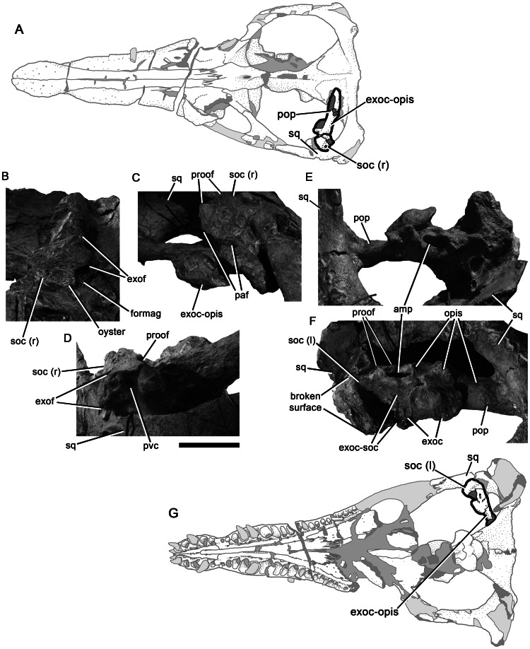
Figure 13. Bones of the otic capsule of Pliosaurus kevani n. sp. DORCM G.13,675.
Schematic (A) showing the portions of the otic capsule figured in (B–D). Right portion of the supraoccipital in posterior (B), right ventrolateral (C) and right posterolateral (D) views. Schematic (G) showing the portions of the otic capsule figured in (E–F). Left exoccipital-opisthotic and articulated left portion of the supraoccipital in anteromedial (E) and ventral (F) views. Abbreviations: amp, ampullary recess in opisthotic; exoc, exoccipital; exoc-opis, exoccipital-opisthotic; exoc-soc, exoccipital-supraoccipital contact; exof, exoccipital facet of the supraoccipital; formag, supraoccipital portion of the foramen magnum; opis, opisthotic; paf, parietal facet of the supraoccipital; pop, parocipital process of the opisthotic; proof, prootic facet; pvc, posterior vertical canal; soc (l), left portion of the supraoccipital; soc (r), right portion of the supraoccipital; sq, squamosal. Scale bar equals 100 mm.

