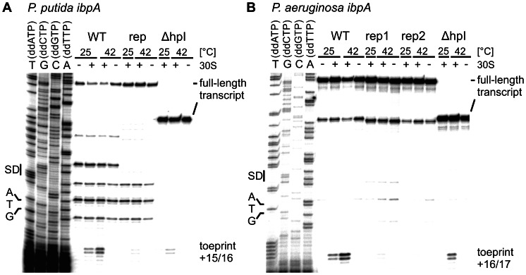
Figure 5. Temperature-dependent binding of the 30S ribosomal subunit to the ibpA 5′UTRs in vitro.
Ribosome binding was shown by toeprinting experiments. The absence (−) or presence (+) of 30S subunits and incubation temperatures are indicated above the gels. Ribosome binding sites (SD) and the ATG start codons are marked to the left of the corresponding DNA sequencing ladder. Full-length products and terminated reverse transcription products (toeprints) relative to the A of the translational start codon are given on the right. A. Toeprinting analysis of P. putida ibpA wt, repressed (U38C/ΔG39) and Δhairpin I (ΔhpI) variants. B. Toeprinting analysis with P. aeruginosa wt, the repressed (rep1 ΔG39; rep2 ΔA35) and ΔhairpinI (ΔhpI) variants.

