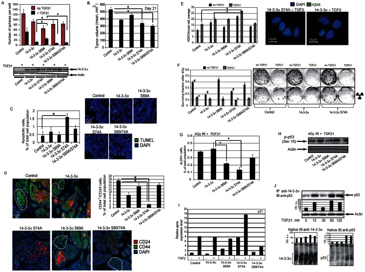
Figure 6. TGFβ1-dependent 14-3-3σ phosphorylation plays a role in regulation of tumor progenitor population.
(A) The role of TGFβ1-dependent 14-3-3σ phosphorylation on TGFβ1-mediated inhibition of sphere initiating population in MCF7 cells. MCF7 cells were stably transfected with the constructs expressing wild type or mutated 14-3-3σ proteins, along with control vector, and used for sphere formation assays in the presence of TGFβ1 at concentration of 5 ng/ml; * - p-value<0.05. (B) Tumorigenic properties of MCF7 cells stably expressing wild type or mutated 14-3-3σ proteins, along with control vector, were analyzed in vivo using NOD/SCID mouse xenograft model. Each experimental group contained at least five mice. Graph shows tumor volumes at day 21 after cell injection; * - p value<0.05. (C) Analysis of cell apoptosis in the xenograft tumors using TUNEL staining. Cells in at least 3 randomly selected fields of view were counted for each condition; * - p-value<0.05. (D) Representative fluorescent images of CD44 and CD24 co-immunostaining in MCF7 xenograft tumors. The percentage of CD44+/CD24− tumor progenitor cells (outlined in white) was counted in at least 3 randomly selected fields of view for each condition; * - p-value<0.05. Scale bar, 30 µm. (E) Immunofluorescence detection of phosphorylated γ-H2A.X at 4 hours after irradiation. MCF7 cells were stably transfected with the constructs expressing wild type or mutated 14-3-3σ proteins, along with control vector, pre-treated with TGFβ1 at concentration of 5 ng/ml for 12 hours and irradiated with 4 Gy X-ray dose. For quantification, at least 100 cells per condition were counted; * - p-value<0.05. Scale bar, 10 µm. (F) Clonogenic radiation survival assay. MCF7 cells were stably transfected with the constructs expressing wild type or mutated 14-3-3σ proteins, along with control vector, pre-treated with TGFβ1 at concentration of 5 ng/ml for 12 hours and irradiated with 4 Gy X-ray dose; * - p-value<0.05. (G) Flow cytometry analysis of tumor progenitor population using ALDEFLUOR assay. MCF7 cells were stably transfected with constructs expressing wild type or mutated 14-3-3σ proteins, along with control vector, treated with TGFβ1 at concentration of 5 ng/ml for 7 days, irradiated with 4 Gy X-ray dose, and analyzed 3 days later; * - p-value<0.05. Representative experiments out of 3 performed are shown. (H) Western blot analysis of p53 phosphorylation at Ser15 in MCF7 cells stably transfected with constructs expressing wild type or mutated 14-3-3σ proteins, along with control vector, treated with TGFβ1 at concentration of 5 ng/ml for 12 h and irradiated with 4 Gy X-ray dose. The cells were analyzed 4 hours after irradiation. (I) Results of semi-quantitative RT-PCR analysis for p21 gene expression in MCF7 cells stably transfected with DNA constructs encoding wild type or mutated 14-3-3σ proteins, along with control DNA plasmid and treated with TGFβ1 at concentration of 5 ng/ml for 12 h. (J) Treatment of the cells with TGFβ1 modulates interaction between endogenous p53 and 14-3-3σ in time dependent manner. MCF7 cells were treated with TGFβ1 at concentration of 5 ng/ml for the indicated times. Cell lysates were immunoprecipitated with anti-14-3-3σ antibody. Anti-p53 antibody was used for immunoblot analysis. The results of blue native PAGE are shown below.

