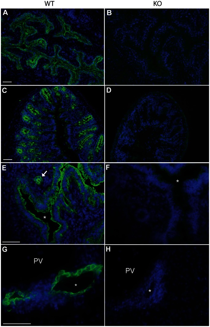
Figure 2. Loss of K7 expression in K7 knockout mouse tissues.
Immunofluorescence microscopy of frozen sections of bladder (A,B), colon (C,D), uterus (E, F) and liver (G, H) of wildtype (A, C, E, G) and homozygous K7 knockout mice (B, D, F, H) stained with polyclonal antibodies to K7 (green). Nuclei are counterstained with DAPI (blue). The asterisks in panels E and F denote the lumen of the uterus. PV denotes the portal vein of the liver; the asterisks in panels G and H indicate the lumen of the bile duct. Scale bars = 50 µm.

