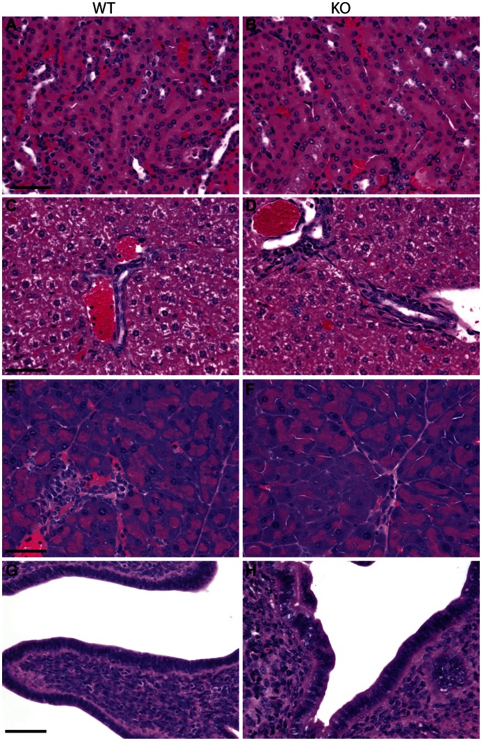
Figure 3. Histological analysis of K7 knockout tissues.
Haematoxylin and eosin stained formalin-fixed tissue sections from wildtype (A, C, E, G) and homozygous K7 knockout mice (B, D, F, H). Images show the cortical collecting tubules of kidney (A, B), bile ducts in liver (C, D), pancreatic ducts (E, F) and columnar epithelium of uterus (G, H). Scale bars = 50 µm.

