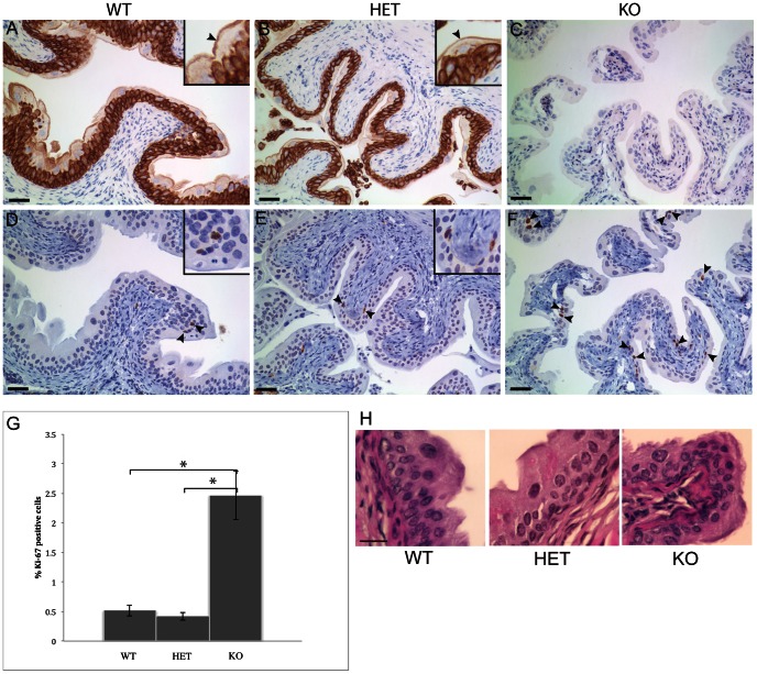
Figure 4. Loss of K7 is associated with hyperproliferation but not hyperplasia of the bladder urothelium.
Immunohistochemistry of bladder sections from wildtype (A, D), heterozygous (B, E) and homozygous K7 knockout mice (C, F) stained with a rabbit polyclonal antibody to K7 (A, B, C) and mouse monoclonal antibody MM1 to the cell proliferation marker Ki-67 (D, E, F). Arrowheads and insets in panels D and E indicate Ki-67 positive nuclei in wildtype (D) and heterozygous K7 knockout (E) bladder. More Ki-67 positive cell nuclei can be seen in the bladder of homozygous K7 knockout mice (arrowheads in F). Scale bars = 50 µm. G. Graph showing the percentage of Ki-67 positive urothelial cells in wildtype, heterozygous and homozygous K7 knockout mice (5 bladders per genotype). For each bladder, 10 random images were collected and an average of 1480 (SD +/−300) urothelial cell nuclei were counted. Standard errors (SE) are indicated by the capped lines. * indicates a p value of less than 0.05 (WT p = 0.01; HET p = 0.007). H. H&E stained sections of the bladder urothelium of wildtype, heterozygous and homozygous K7 knockout mice. Scale bar = 25 µm.

