
Figure 8. Cd regulated the expression levels of Bcl-2 and Bax.
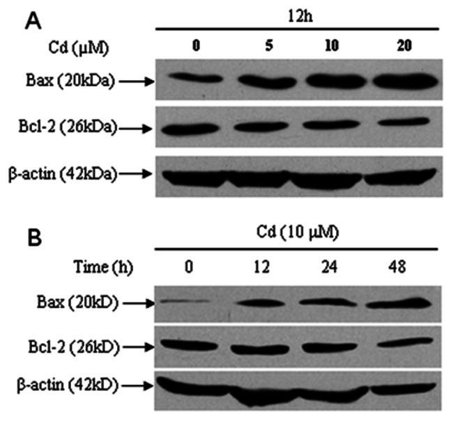
(A and B) Cerebral cortical neurons were treated with Cd (0, 5, 10, 20 µM) for 12 h, or 10 µM Cd for 0–48 h and then assessed by Western blotting. β-actin was used as an internal control. The expression level of Bcl-2 decreased, while that of Bax increased. All experiments were performed twice.