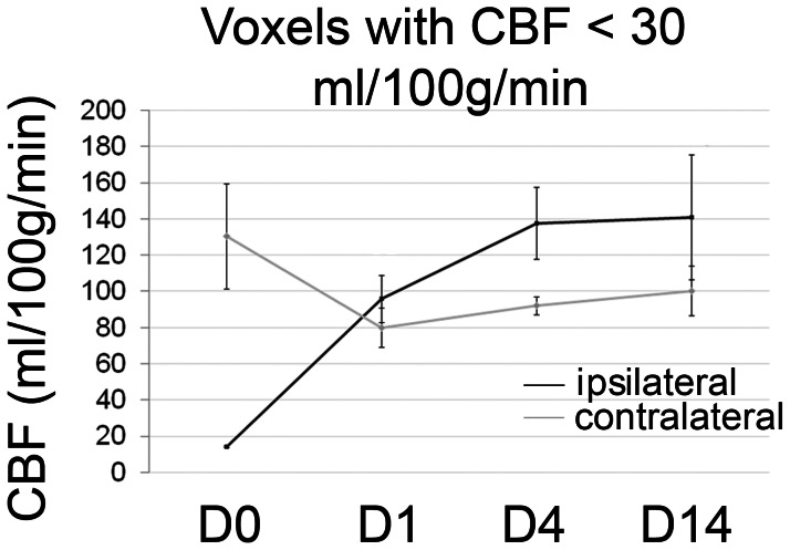
Figure 2. CBF in ischemic voxels over time.
Black line: CBF changes within voxels with intra-ischemic CBF of <30 ml/100 g/min between d 0 (D0; during vascular occlusion) up to d14 after MCAO. Grey line: contralateral CBF values from the same animals. Mean ± SD values are shown. The GLM analysis for repeated measures demonstrated a significant effect of the within-subject factor “time” (p = 0.03), but not of the between-subject factor ipsi- or contralateral side (p = 0.7), but for the interception of the two factors (p<0.0001).

