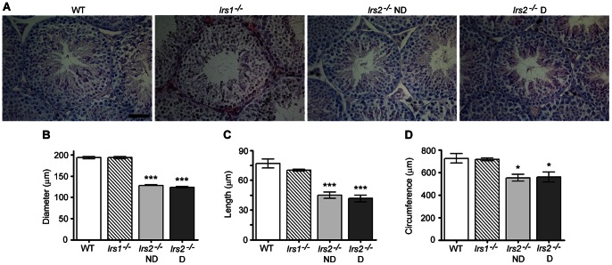
Figure 2. Testis morphology of Irs2 −/− mice is normal but structures are reduced in size.
Transverse histological sections of 5 µm were cut through the long axis of Bouin's-fixed paraffin-embedded testes. (A) Hematoxylin and eosin staining of testicular cross sections demonstrates normal cellular associations in the seminiferous epithelium and numerous elongated spermatozoa extending into the lumen. Representative images were captured using a 40x objective. The scale bar represents 50 µm. (B) The diameter of the seminiferous tubules, (C) the length of the seminiferous epithelium, and (D) the circumference of the seminiferous tubules were reduced in Irs2-deficient but not in Irs1-deficient mice. Results for each measurement are mean ± SEM from a minimum of 50 randomly selected seminiferous tubules from five mice from each phenotype. Asterisks denote a significant difference compared to WT; * P<0.05, *** P<0.001. Adult mice of 8–12 weeks of age were used for the study.

