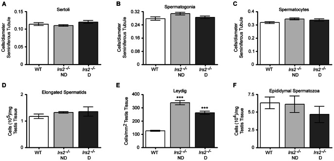
Figure 5. Irs2-deficient mice have reduced testicular cell number but normal cell density.
For (A) Sertoli cells, (B) spermatogonia, and (C) spermatocytes, the number of cells per cross section of seminiferous tubule was normalized to the diameter of the seminiferous tubule. There were no differences between WT and Irs2-deficient mice (P>0.05). (D) The number of elongated spermatids was normalized to the weight of testis parenchyma. No significant differences were observed between WT and Irs2-deficient mice (P>0.05). (E) The number of Leydig cells was normalized to square area of testis tissue and there were significantly more Leydig cells per equivalent square area in Irs2-deficient testes compared to WT animals (P<0.001). (F) The number of spermatozoa collected from epididymides was normalized to testes weights. There were no significant differences between WT and Irs2-deficient mice (P>0.05).

