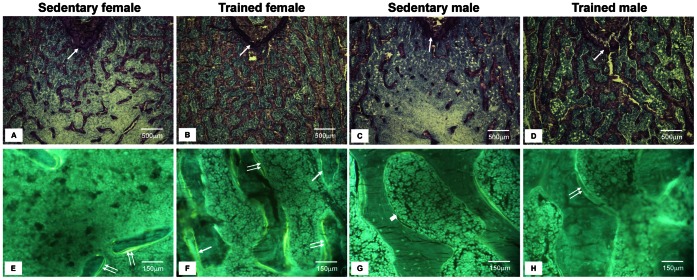
Figure 1. Illustrative bone histological characteristics of sedentary and trained animals.
1A–1D. Undecalcified Bone: Characteristic light microscopy aspects of trabecular bone (femoral metaphysis). Toluidine blue staining showing an increase in the trabecular bone volume (BV/TV) and trabecular thickness (Tb.Th) in the trained animals (B and D) compared with their sedentary counterparts (A and C). The epiphyseal growth plate is indicated by arrows. Histomorphometric analyses were performed at 195 µm under the epiphyseal growth plate (Magnification, x40). 1E–1H. Double oxytetracycline labeling: Characteristic fluorescent light microscopy of undecalcified bone (femoral metaphysis). Unstained bone sections under UV light of the sedentary (E and G) and trained (F and H) animals. Single and double labels are indicated by the single and double arrows, respectively. By quantifying the distance between the oxytetracycline double-labels, we observed that the trained males (H) presented a greater mineral apposition rate (MAR) than the sedentary males (G) and trained females (F). By evaluating the percentage of the trabecular bone surface that was double-labeled, we calculated the bone formation rate (BFR/BS), which was increased only in the trained males (H) (Magnification, x250). Details of the histomorphometric results can be found in Table 4.

