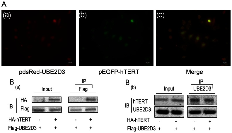
Figure 2. hTERT interacts with UBE2D3 proteins.
(A)pEGFP-hTERT and pdsRed-UBE2D3 were co-transfected into MCF-7 cells. UBE2D3 with red fluorescent tag(A-a), hTERT with green fluorescent tag(A-b). The 3rd pic is the merge(A-c). From the picture above, we could indicate most of them express in the nucleus and the possibility of interaction between them in space. (B-a)HA-tagged hTERT and/or FLAG-tagged UBE2D3 plasmids were co-transfected into HEK293T cells as indicated. At 24 h after transfection, whole-cell lysates were isolated. Cell lysates were immunoprecipitated with anti-FLAG and the hTERT proteins in the complex identified with immunoblotting with anti-HA (top panel). UBE2D3 and hTERT protein expression was demonstrated with direct immunoblotting(IB) of cell lysates with HA antibody and FLAG antibody (left panel or input), respectively. (B-b)The above plasmids cotransfected into HEK293T cells as indicated. Cell lysates were immunoprecipitated with UBE2D3 and the hTERT proteins in the complex identified with immunoblotting with anti-hTERT (top panel). UBE2D3 and hTERT protein expression was demonstrated with direct immunoblotting of cell lysates with UBE2D3 and hTERT antibody (left panel or input), respectively. All experiments were repeated 3 times with similar results.

