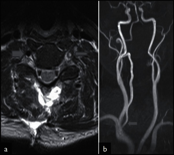
Figure 1.

MRI detection of vertebral artery injury. Axial T2-weighted MRI (TE 91 msec; TR 4100 msec; (a) shows hyperintensity in the region of left vertebral artery flow void (thin arrow). The right vertebral artery shows normal flow void (thick arrow). Coronal maximal intensity projection (MIP) of TOF angiography (b) reveals the absent left vertebral artery. The right vertebral artery shows normal flow related enhancement (arrow)
