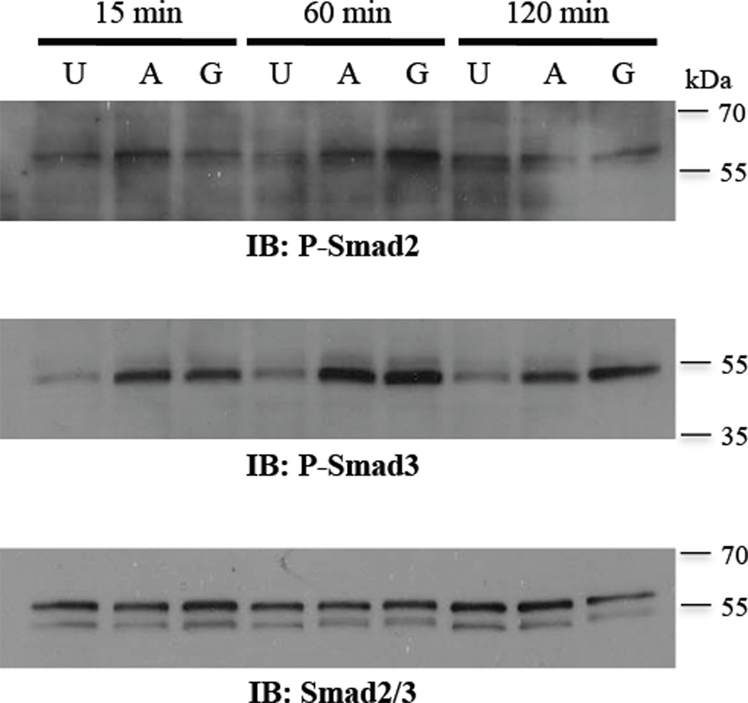
Figure 4. Smad phosphorylation by activin and GDF-9 in primary rat GCs.
Treatment of GCs with 100 ng/ml activin A (A) or 300 ng/ml GDF-9 (G) increases phosphorylated Smad2 (P-Smad2) and Smad3 (P-Smad3) within 15 min with maximal activation at 60 min, compared with untreated (U) cells. For total Smad2/3, the antibody detected both total Smad2 (upper band, 60 kDa) and Smad3 (lower band, 52 kDa).

