Figure 2.

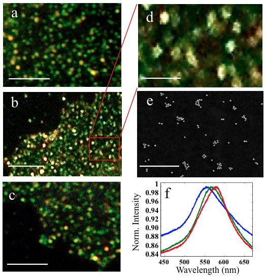
Au NP immunolabels form discrete spots on the cell membrane for different cell/protein targets: a) SKBR3/CD24, b) MCF7/CD24, c) MCF7/CD44. The magnified view in d) further emphasizes the “spotting” after labeling with NPs. e) SEM imaging confirms the organization of the cell-bound NPs into clusters of varying sizes (here for MCF7/CD24). f) Exemplary spectra of individual spots on the cell surface. Size bars are 5μm for a–c), 1μm for d–e).
