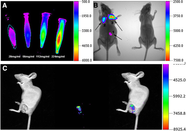
Figure 6.
AuNCs@SiO2-FA nanoprobes for fluorescence imaging. (A) In vitro fluorescence image of AuNCs@SiO2–FA in 0.01 M PBS with different concentrations; (B) In vivo fluorescence image of 50ul AuNCs@SiO2-FA injected subcutaneously at three different dose (area-1: 56 mg/ml; area-2: 112 mg/ml; area-3: 226 mg/ml) into the left mice. The left mice without injected was selected as control; (C) Fluorescence image of tumor tissues with tail vein injection at the concentration of 287 mg/ml at 6 h post-injection.

