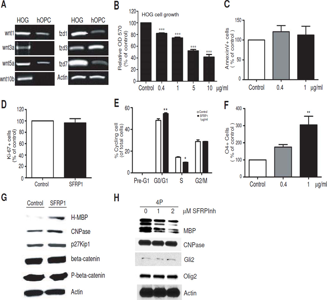
Figure 2.
Inhibition of Wnt signaling with recombinant SFRP1 causes HOG cell growth arrest and differentiation. A. Semi-quantitative PCR analysis showing HOG cells after 3 days in culture express transcripts for Wnt ligands and Frizzled receptor forms. B. SFRP1 retards HOG cell growth. HOG cells were incubated with the indicated concentrations of recombinant SFRP1 for 3 days and MTT assays were performed. All values shown are mean ± SEM of at least 3 independent experiments. ***P<0.001, one way ANOVA, Dunn’s posthoc test. C. Annexin V assay reveals no significant difference in cell survival after treatment with SFRP1 for 3 days. Values are expressed as percentage over control samples. D. No significant change in the percentage of Ki-67+ cells is observed after 3 days of treatment with 1ug/ml SFRP1. Values are expressed as percent Ki-67 positive cells of a normalized total of sorted events. E. Cell cycle analysis of HOG cells treated with 1ug/ml SFRP1 for 3 days. Propidium iodide staining reveals reduction of cells in S phase accompanied by accumulation in G0/G1. Histograms from a representative experiment is shown. Values are expressed as a percentage of total cells analyzed. * P<0.05, ** P<0.01, vs control, Student’s T-test. F. SFRP1 treatment increases HOG differentiation to the O4 stage. FACS-assisted quantitative analysis of O4+ cells following treatment with increasing doses of SFRP1 for 3 days. Values are percentage O4+ cells of total sorted events further expressed as percentage of control. ** P<0.01, one way ANOVA, Dunn’s posthoc test. Values shown in histogram panels are mean ± SEM of at least 3 independent experiments. G. Western blot analysis of proteins in HOG cells shows increased expression of H-MBP, CNPase and p27 after treatment with 1 ug/ml SFRP1 for 3 days. H. Western blot showing myelin protein response of primary rat OPs to SFRP1 inhibitor (SFRPInh). Rat OPs were cultured in the presence of 10 ng/ml human recombinant PDGF-AB for 4 days (4P) in the absence or presence of SFRP1 inhibitor at the indicated concentrations. Control samples received an equal volume of DMSO.

